Fungi in Sandall Beat Wood December 2023
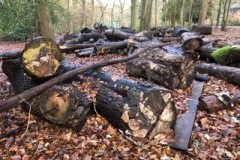
Near to the Visitor’s Centre there is a pile of logs many of which have been there for several years
and display an amazing array of fungi including brackets , rots, crusts and slime moulds. Most of
the logs are beech, oak or silver birch. Here is a photo of the site with the Visitor’s centre just
showing in the top right hand corner of the photograph.
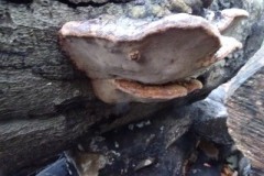
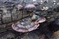
The following two photographs taken from above and from the side are examples of a species of
Ganoderma.
Several fungi included in this Blog have been identified by Kevin Gilfedder but only from a
photograph so in some cases the identification is only a probability. Kevin pointed out that extra
pieces of information as well as a photograph are helpful when attempting an identification. They
include: the substrate on which the fungus is growing, a photograph of the underside, whether or
not the flesh bruises when pressed and if so what colour and any particular smell.
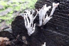
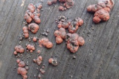
Here are two more examples. The first an easily recognisable fungus called Candlesnuff Xylaria
hypoxylon which is very common and found on broad-leaved trees and the second a red slime
mould called Lycogala epidendron in its fruiting stage ready to shed spores.( identified by Kevin )
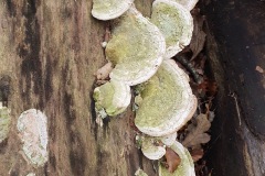
Kevin directed me to the genus Trametes for this next fungus and initially we weren’t sure whether
it was Trametes pubescens or Trametes gibbosa , which is more common than the former.
Apparently many of the brackets develop algae on their upper surface so that feature wasn’t very
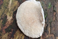
useful in determining which species it was. Fortunately I took Kevin’s advice and photographed
the underside which clearly showed the irregular, elongated maze-like pores which distinguish
gibbosa from other species of Trametes which have round or oval pores.
Nora
Robin’s Pincushion Diplolepsis rosae – a closer look
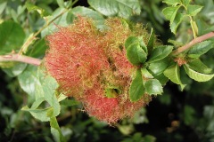
Robin’s Pincushion or the Bedeguar gall is induced by the cynapid ( gall ) wasp Diplolepsis rosae. In Germany it is known as the”sleep apple” as it is said to aid sleep if placed under one’s pillow. This strikingly beautiful gall, which incidentally is the emblem of the British Plant Gall Society, is found on species of wild rose, mainly Rosa canina. This photograph, taken in late July, shows a gall at its most attractive stage. The mass of reddish branched filamentous growths contain vascular strands which are continuous with those of the plant’s vascular system. (S. Randolph).
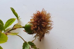
The life of the gall starts in early spring when a female wasp lays its eggs, usually in buds, but as the next photograph shows, occasionally on hips.
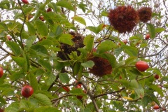
Often several galls develop on the same bush, as you can see in the next photograph.
Starting as early as late April, the female gall wasp taps the surface tissues of buds with her antennae until a suitable expanding bud is found. This stage could take as long as 60 minutes. Then with her head facing downwards she uses her long curved ovipositor to probe the surface of
the bud to locate the most suitable position for insertion of eggs. Once in position she will insert her ovipositor with remarkable precision under the edge of an outer bud scale and remain motionless for 1-2 hours, during which she can lay 30- 100 eggs in the same bud. Each egg is glued to a single epidermal cell. Cell proliferation in the area of oviposition begins before the egg hatches in order to create a cavity into which the larva crawls once the egg has hatched, which will be within a week of oviposition. The eggs are produced by a method of reproduction called parthenogenesis which means that reproduction occurs without the necessity for fertilisation. It is likely that a single
female will lay eggs in more than one bud. Synchronisation between the emergence of the egg laying females and the expansion of the buds on the host plant is essential. Initiation of the gall itself arises as the larva starts to feed, causing mechanical damage to the cells around it with its mandibles. Several other factors are thought also to be responsible, including the wounding of tissue by oviposition. The larva becomes surrounded by an inner chamber around which layers of nutritive cells develop. These cells become layers which are continuously replaced as the larva feeds and are found nowhere else in the plant, only as part of the structure of the gall. Through the cells the gall causer stimulates the plant to send vast amounts of nutrients from other parts of the plant through a network of vascular bundles. Around the inner chamber a layer of thick walled cells develop protecting the inner chamber. The fully developed Robin’s Pincushion consists of many chambers each housing an inducer larva, with the outer thick walled layers coalescing and surrounded by a mass of branched sticky filaments.These hairy filaments are initially green-pink but turn brown to black in Autumn/Winter. A gall with many chambers like this is an example of a multiocular gall. During the period of rapid development of the gall, (the gall tissue is fully formed and reaches maturity within four to eight weeks ) the larva feeds only slowly and undergoes a series of moults until it is fully developed in late October. It then ceases feeding and enters the prepupal stage during which it overwinters. Pupation occurs inside the chambers the following March or April and
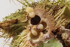
the final development of the adult takes only a short time. The adult cynapid wasps emerge in April using their strong mandibles to chew exit holes in the gall. The exit holes can be seen more easily when the filamentous branches fall away. The majority are females capable of laying fertile eggs without mating.
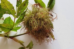
The following photograph, taken in August, shows the outside of a fully formed gall.
The next photograph of the same gall cut in half shows several chambers, the hard tissue around them, several larvae most probably the gall causing larvae and the pink and green filamentous growths.
The number of cells or chambers within a single gall varies but there can be as many as 60 chambers. Smaller galls may have as little as 2 chambers, as you can see in the following photograph.
The size of the gall depends upon three factors, the number of inner galls inside, the choice of bud, a terminal bud receiving more of the plants resources and so growing more vigorously can support more larvae and on the survival of the larvae inside. Should the larvae die after the gall has started to grow the inner galls may not develop any further but the hairs on the outside continue to grow and the result is a solid gall covered in the hair-like filaments. Robin’s Pin cushions with their many chambers filled with plump larvae are attractive to other insects which seek to take advantage of the food and shelter to be found in the galls. These insects are either inquilines , a term which means guests, which lay their eggs in the gall with the intention of using the same food source as the host and may or may not result in the death of the gall causer, and parasitoids which deliberately lay their eggs either on the surface ( ectoparasitoids ) or in the body (endoparasitoids ) of the inducer larvae. Both types paralyse their host larva by injecting venom and then feed on the living body until it dies.
The one known inquiline which uses Robin’s Pincushion is another cynapid wasp called Periclistus brandtii. An assortment of parasitoids are known to feed on the host larvae. Since their action always results in the death of the gall causer, they are parasitic. Together with the host
inducer they form a community. During the winter the galls lose most of their hairs and so once the chambers are vacated the holes are more obvious and a variety of creatures take advantage of the gall as shelter. These, known as successori, use the old empty cells as a place to rest or hibernate and include moths, earwigs bugs etc.They may also feed on fungi and bacteria that colonise the spaces.
References.
Britain’s Plant Galls by Michael Chinery.
The Natural History of the Rose Bedeguar Gall and its insect community by S. Randolph.
The Natural History of the Rose Bedeguar Gall part 2 by Robin Williams
Plant Galls by Margaret Redfern
Crown galls
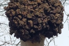
Crown gall causing bacteria Agrobacterium humefaciens infect plants by inserting some of their own DNA into plant cells through a wound. This alters their function causing them to produce unusual amounts of plant hormones which in turn stimulate the plant cells to multiply and enlarge leading to the tumour formation. The bacterium forces the plant to produce opines which it uses for energy. Even if the bacterial infection is cleared gall growth continues.
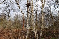
These galls are quite common on Silver Birch tree trunks. Both photographs below taken in Park Wood, Rossington. The first by Nora Boyle and the second by Caroline Barrass.
